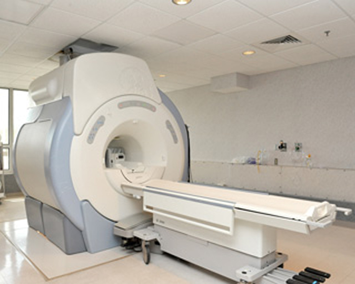
Why an MRI?
Due to fine detail capabilities of MR images compared to standard X-rays, ultrasounds and CT scans, it is used to find serious problems such as tumors, bleeding, injury or infection. It may also be used to further examine a problem that was found on an X-ray, ultrasound or CT scan. An MRI scan may be done on the following areas of the body:
- Head – An MRI can look for tumors, aneurysms, bleeding in the brain, or damage caused by a stroke. It can also be used to find problems of the eyes and ears.
- Chest – An MRI can look at the heart, valves and coronary blood vessels and to see if there is any damage to the heart or lungs. It can also look for breast cancer and lung cancer.
- Blood Vessels – MRIs can locate problems of the arteries and veins, such as an aneurysm, blocked blood vessel or a torn lining of a blood vessel.
- Abdomen and Pelvis – An MRI can identify problems in organs such as the liver, gallbladder, pancreas, kidneys and bladder. It can also be used to view the uterus and ovaries in women and the prostate in men.
- Bones and Joints – MRIs can check for problems such as arthritis, bone marrow problems, bone tumors, cartilage problems, torn ligaments or tendons, or infection. Also, an MRI may be used for further review if a standard X-ray was unable to determine if a bone is broken or not.
- Spine – MRIs can check the discs and nerves for spinal stenosis, disc bulges and spinal tumors.
How to Prepare
As with any health care procedure, being prepared plays an important part in the outcome of your procedure and your satisfaction as a patient. To help you prepare for your MRI we have a few tips. First of all, tell your doctor or MRI technologists if you:
- Are allergic to any medications
- Are or might be pregnant
- Have a pacemaker, artificial limb, any metal pins or parts in your body, metal heart valves, metal clips in your brain, metal implants in your ears, tattooed eyeliner or any other implanted or prosthetic medical device
- Have had an accident or work around metal
- Have had recent surgery on a blood vessel
- Have an intrauterine device (IUD) in place
- Become very nervous in confined spaces
- Wear any medicine patches
You will be asked to sign a screening and consent form saying that you understand and agree to the risks of an MRI before you have your procedure done; your doctor will review the potential risks in detail when discussing the test with you. Please talk to your doctor if you have any concerns or questions over this procedure. You may also need to arrange for someone to drive you home after the test if you are given medicine to help you relax.
Please take note: It is also very important to remember to remove all metal objects such as hearing aids, dentures, jewelry, watches and hairpins from your body, as these objects will be attracted to the magnet used for the test.
During Your MRI
During your test, you will lie on your back on a table that is part of the MRI scanner. In order to help you remain still, you may be strapped in at your arms, head, and chest. A special device called a coil may be placed over or wrapped around the area to be scanned and a special belt strap may be used to sense your breathing or heartbeat.
Once the test begins, the table will slide into the space that contains the magnet. Inside the scanner you will hear a fan and feel air moving. You may also hear tapping or snapping noises as the MRI scans are being taken. It is very important that you hold completely still while this is going on to ensure clear and accurate images are taken. The test should last 30 minutes to an hour, although some can take as long as two hours.
We understand that an MRI can be an uncomfortable experience and can cause some uneasiness, especially if you feel nervous in tight spaces. To help ease your discomfort, we’ve located our MRI in a well-lit room with large windows, to allow plenty of natural light. Your doctor may also be able to give you a sedative if your nervousness prevents you from holding still. Please discuss this with your doctor prior to your MRI appointment.
There is no pain involved during your MRI test and no known harmful effects from the powerful magnetic field used for imaging. The radiologist will review the findings and send a report to your physician who will be able to discuss the complete results with you.
If you have questions about an upcoming MRI, please contact our Diagnostic Imaging staff by calling 740-623-4132. For more information on Diagnostic Imaging testing and treatments, visit RadiologyInfo.org.
Featured Services

Surgical Clinic

Emergency Services
Urgent Care
Physical and Occupational Rehabilitation


