We also recommend that:
- You do not wear deodorant, talcum powder or lotion under your arms or breasts, as those substances can appear on the mammogram as calcium spots.
- Describe any breast symptoms or problems to the technologist performing the exam.
- If you can, obtain prior mammograms and make them available to the radiologist at the time of your exam.
- Ask when your results will be available, and check with your physician for the results. Don’t assume the results are normal if you don’t hear anything back.
During Your Digital Mammogram
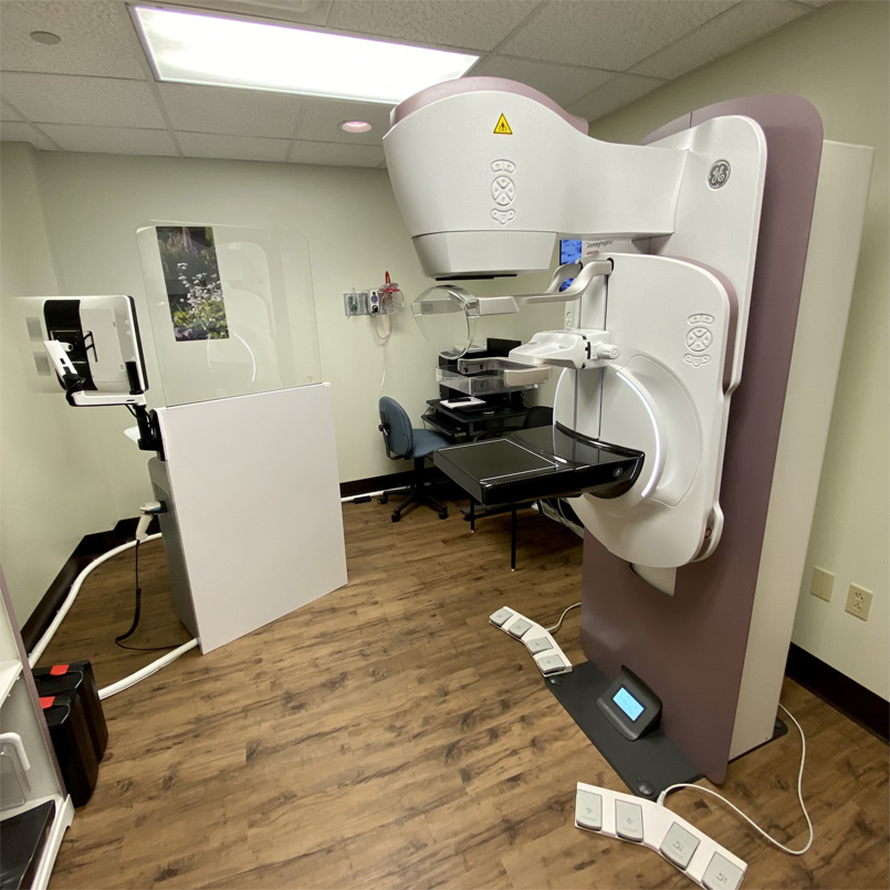
A digital mammogram is performed almost exactly the same way a conventional film screening mammogram is. During your procedure, a radiologist will position your breast in the mammography unit. Your breast will be placed on a platform and compressed with a paddle. The technologist will gradually compress your breast. This compression is necessary in order to:
- Even out the thickness of the breast so a clear image of all of the tissue can be taken
- Spread out the tissue so that small abnormalities can be found
- Allow the use of a lower X-ray dose since a thinner amount of breast is being manages
- Hold the breast still in order to avoid blurring the image
- Reduce X-ray scatter to increases the clarity of the picture
The technologist will ask you to change positions between capturing images and you must hold very still and may be asked to hold your breath while the picture is being taken. The routine views are top-to-bottom and an oblique side view.
You will feel a slight pressure on your breast during the exam, and for women with sensitive breasts, this can cause discomfort. Be sure to inform the technologist if the pain increases as the compression does, as less compression can be used to make you more comfortable.
The exam should last about 30 minutes and your results will be sent to your physician after the radiologist has analyzed them.
Featured Services

Surgical Clinic

Emergency Services
Urgent Care
Physical and Occupational Rehabilitation


